Ear Anatomy
The human ear is the organ of hearing that detects, analyzes sound by transduction (or the conversion of sound waves into electrochemical impulses) and maintains the sensation of balance. The anatomy of the ear is divided into outer, middle, and inner ear.
Thus, this organ allows us to listen. Hearing can be defined as the perception of sound energy through the brain and central nervous system. This energy passes through the different parts of the ear anatomy as explained below.
Outer ear
The outer ear is the part that acts as a funnel to conduct air vibrations to the eardrum. It also has the function of sound localization. The outer ear is made up of the pinna and the ear canal.
Pinna
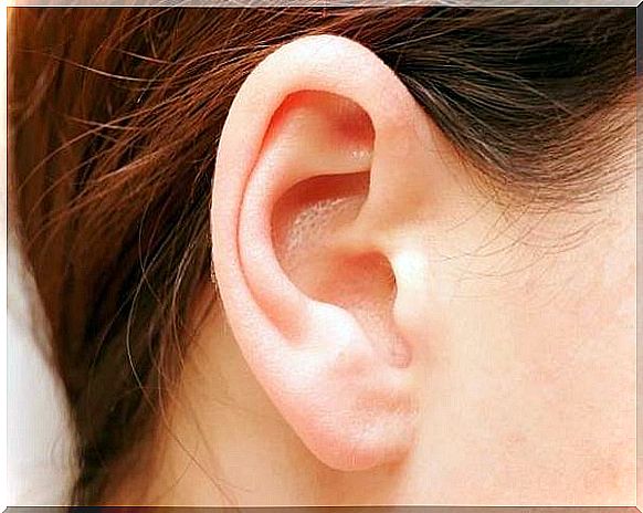
Within the anatomy of the ear is the outermost part. Thus, it is a prominent flap covered by the skin that is located on the sides of the head, and is the externally visible part of the ear. It is formed and supported by cartilage except for the earlobe.
It collects sound waves and channels them into the external auditory canal through patterns formed in the pinna known as whorls and hollows.
Its shape also partially shields sound waves approaching the ear from the back, allowing a person to know whether a sound is coming directly from the front or not.
Ear canal
It is approximately three centimeters long in adults. It is supported by cartilage at the opening, and by bone for the remainder of its length. The skin lines the canal and contains glands that produce secretions that mix with dead skin cells to produce earwax.
Earwax, along with the fine hairs that protect the entrance to the ear canal, help prevent airborne particles from reaching the inner parts of the ear canal.
Earwax usually dries up and falls out of the canal. However, it can sometimes become compact and disrupt hearing.
Middle ear
The middle ear is located between the outer and inner ear. It is separated from the ear canal from the outer ear by the tympanic membrane (the eardrum).
It works by transferring vibrations from the eardrum to the fluid in the inner ear. This transfer of sound vibrations is possible through a chain of small mobile bones, called ossicles , which extend through the middle ear and the corresponding small muscles.
Tympanic membrane (eardrum)
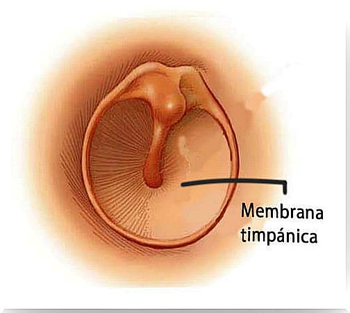 The tympanic membrane is commonly known as the eardrum and separates the ear canal from the middle ear. Some of its characteristics are:
- It is slightly concave.
- It vibrates freely in response to sound.
- It measures about 1 centimeter in diameter.
- The membrane is highly innervated, making it very sensitive to pain.
- For the membrane to move freely when air hits it, the pressure of the resting air on both sides of the tympanic membrane must be equal.
The exterior is exposed to atmospheric pressure through the auditory tube, so that the cavity in which it is located, called the tympanic cavity, is continuous with the cells in the jaw.
Normally, the auditory tube is elongated and closed, but by swallowing, yawning and chewing, it opens, allowing air to enter or leave the tympanic cavity. This opening of the auditory tube allows the air pressure in the middle ear to balance with atmospheric pressure.
Auditory ossicles and muscles
The tympanic cavity contains the smallest bones and muscles of the body. From the outermost to the innermost, they are:
- Hammer. Attached to the eardrum and has a handle that attaches to the inner surface of the eardrum. It also consists of a head suspended in the tympanic cavity.
- Anvil. It is connected to the hammer and stirrup.
- Stirrup. It consists of an arch and a platform. It is held by a piece of ring tissue in an opening called the oval window, which is the entrance to the inner ear.
What converts the sound wave vibrations into the movement of the inner ear fluid is the tensor tympani, the muscle of the inner ear that is attached to the hammer.
Inner ear
Within the anatomy of the ear, the inner ear is the deepest part of the entire ear, and is located in a place known as the bony labyrinth.
A cushion of fluid, called a perilymph , lies between the bony labyrinth and the membranous, while a fluid called endolymph is found within the membranous labyrinth itself.
Inside the inner ear there is a chamber called the vestibule, which plays an important role in the sense of balance.
Cochlea
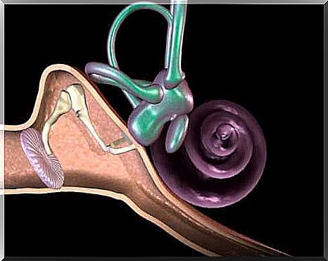
From the vestibule comes the cochlea, sometimes referred to as: the organ of hearing. It is the part of the whole ear that actually converts sound vibrations into auditory perception.
- It is shaped like a spiral in the shape of a snail.
- It is about 9mm wide at the base and 5mm high.
- It wraps around a section of cancellous bone called a modiolus.
- Inside is the organ of Corti, with hair cells that transform sound vibrations to reach the brain.
Cochlea chambers
The cochlea contains three fluid-filled chambers separated by membranes:
- The upper chamber or vestibular ramp.
- The scala lobby or middle ramp.
- The lower chamber or scala tympani.
The scala vestibuli is separated from the scala media by Reissner’s membrane and the scala media is separated from the scala tympani by the basilar membrane, which is where the organ of Corti is located.
Organ of Corti
The organ of Corti is supported by a membrane called the basilar membrane . It is about the size of a pea, and acts like a transducer, converting vibration into nerve impulses. That is, it transforms the mechanical energy of sound into nervous energy that reaches the brain. It consists of:
- Support cells.
- Hair cells, which have long, rigid microvilli called stereocilia on the apical surfaces.
Microvilli are fine hair-like structures in cells that help increase cell surface area. In addition to these stereocilia, it has a gelatinous membrane called the tectorial membrane. Four rows of hair cells rotate along the organ of Corti.
These outer hair cells adjust the response of the cochlea to different sound frequencies to allow the inner hair cells to function more precisely.
Since it never hurts to know how the body works, now we have taught you how the ear works and the sense of hearing that we value so much. Without a doubt, the anatomy of the ear makes it possible for humans to hear the sounds of the world around them.